PAPILLON-LEFEVRE SYNDROME AND PERIODONTAL DISEASE
Description
Periodontitis: Inflammatory disease of the supporting structures of the periodontium including the gingiva, periodontal ligament, bone and cementum. This condition shows irreversible destruction to tissues resulting in bone loss.
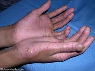
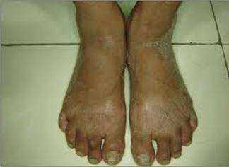
Papillon-LeFevre Syndrome: Rare autosomal recessive trait characterized by palmar plantar hyperkeratosis. The soles of the feet as well exhibit this disorder. Intraorally there is periodontal destruction and premature exfoliation of the deciduous and permanent dentition. Also known as palmoplantar keratoderma.
Papillon-LeFevre Syndrome: Rare autosomal recessive trait characterized by palmar plantar hyperkeratosis. The soles of the feet as well exhibit this disorder. Intraorally there is periodontal destruction and premature exfoliation of the deciduous and permanent dentition. Also known as palmoplantar keratoderma.
Distinguishing diagnostic factors
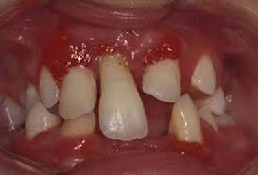
Patients with this condition are normal at birth except for a reddening of the palms and soles of the feet. Teeth erupt at normal sequence, but at around 1 1/2 to 2 yrs a gingivoperiodontal inflammatory process begins. The oral cavity is marked by edema, bleeding, alveolar bone resorption and mobility of teeth, which usually ends up with exfoliation of teeth.
Periodontal effects appear almost immediately after tooth eruption, when the gingiva becomes erythematous and
edematous.
Plaque usually accumulates in the deep crevices. Usully the primary incisors are affected first around 3 yrs of age. It has been found that by 4 or 5 yrs old, all the primary teeth have been exfoliated. Following tooth loss, the gingival appearance resolves and may appear to return to health. However, the process repeats during the permanent dentition and exfoliation completes around 15 years old.
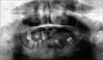
Destruction of bone and mobility of the teeth give a radiographic appearance of "floating teeth."
Periodontal effects appear almost immediately after tooth eruption, when the gingiva becomes erythematous and
edematous.
Plaque usually accumulates in the deep crevices. Usully the primary incisors are affected first around 3 yrs of age. It has been found that by 4 or 5 yrs old, all the primary teeth have been exfoliated. Following tooth loss, the gingival appearance resolves and may appear to return to health. However, the process repeats during the permanent dentition and exfoliation completes around 15 years old.
Destruction of bone and mobility of the teeth give a radiographic appearance of "floating teeth."
Additional symptoms and findings associated with this disease are pyogenic skin infections, abnormalities of the nails, and excessive perspiration.
Etiology of the disease or condition
The gene to this syndrome was found on chromosome 11 regions 14-21. It is passed down as an autosomal recessive gene.
Pathogens associated with the disease or condition
It has been found that deep sub-gingival flora is composed of motile gram negative anaerobic rods, such as Bacteroides gingivalis, Capnocytophaga and a number of spirochetes. Actinobacillus actinomycetem-comitans was also found as being commonly responsible in the pathogenesis of periodontal destruction in these patients as well as P. gingivalis.
Because periodontal destruction occurs, it is believed to be due to the decreased chemotaxis of neutrophils in the periodontium to fight of the bacteria. Research suggests that bacterial and viral involvement are the initiating factors for the periodontal destruction.
Because periodontal destruction occurs, it is believed to be due to the decreased chemotaxis of neutrophils in the periodontium to fight of the bacteria. Research suggests that bacterial and viral involvement are the initiating factors for the periodontal destruction.
Classification of Disease or Condition
AAP Class Type IV: Periodontitis as manifestation of Systemic Disease: Genetic factors
Prevalence of the disease or condition
Usually first appears in childhood and manifests again when the permanent dentition appears.
Factors to Include in Patient Education
Because all form of treatment to date has not been able to prevent the periodontal disease destruction, and exfoliation of teeth, it is important that the patient stay in contact with the dentist and periodontist to monitor the progression of the syndrome. SInce gingival health resumes a normal appearance after, it is important to monitor the extent of bone destruction.
Treatment Recommendation
Conventional periodontal therapy have been unsuccessful in the treatment of this disease. Oral Retinoids and antibiotics have been successful in treating the skin condition associated with this syndrome, but has not helped with the periodontal destruction. To replace missing teeth, it is recommended to have prosthetics or implants placed. A combination of overdentures and implants have been commonly performed to replace missing teeth. Bone grafting is necessary for severe alveolar bone destruction.
Maintenance Recommendation
Frequent monitoring is necessary to update and futher bone loss progression and gingival health.
References:
Vikhe, D. M., Lagdive, S. B., Gangadhar, S. A., & Bhandari, A. A. (2011). Prosthodontic Rehabilitation of Patients with Papillon - Lefevre Syndrome: A Case Report. Pravara Medical Review, 3(4), 19-22.
Ibsen, O.A, Phelan, J.A, Oral Pathology for the Dental Hygienist, 5th edition 2009.
Vikhe, D. M., Lagdive, S. B., Gangadhar, S. A., & Bhandari, A. A. (2011). Prosthodontic Rehabilitation of Patients with Papillon - Lefevre Syndrome: A Case Report. Pravara Medical Review, 3(4), 19-22.
Ibsen, O.A, Phelan, J.A, Oral Pathology for the Dental Hygienist, 5th edition 2009.
All images taken from Google Images
